Bone Cancer refers to malignant tumors that arise in bone tissue, including both primary bone cancers and secondary (metastatic) bone cancer that spread from other organs. Primary bone cancers originate in the bone itself, whereas metastatic disease reflects dissemination of cancer cells from sites such as the breast, prostate, lung, or kidney. Major primary subtypes include osteosarcoma, chondrosarcoma, and Ewing sarcoma, each with distinct age patterns and biological behavior. The underlying mechanism involves uncontrolled growth of abnormal cells that disrupt normal bone remodeling, invade surrounding tissues, and can weaken the structural integrity of bone. Contributing factors may include inherited cancer predisposition syndromes, prior exposure to ionizing radiation, and certain benign bone conditions that predispose to malignant transformation, though many cases occur without a clearly identifiable cause.
Bone cancer commonly presents with persistent bone pain that may worsen over time and can be accompanied by swelling or a palpable mass near the affected bone. Tenderness and localized warmth may occur, and pain can be aggravated by activity or at night, depending on the tumor’s location and aggressiveness. As the lesion expands, it may reduce range of motion in nearby joints and contribute to functional limitations. Some patients present with an increased tendency for pathologic fractures, meaning fractures that occur with minimal trauma due to weakened bone. Symptoms can vary by subtype and site, with Ewing sarcoma often affecting children and adolescents and sometimes producing systemic features such as fatigue or fever, while chondrosarcoma more often occurs in older adults and may progress more slowly.
The concept of bone malignancy has evolved with advances in pathology, imaging, and molecular genetics, enabling clearer separation of tumor types that previously were grouped together. Osteosarcoma, chondrosarcoma, and Ewing sarcoma have long been recognized as distinct entities based on microscopic appearance and clinical behavior, and modern classification increasingly incorporates genetic and molecular markers. Epidemiologically, primary bone cancers are uncommon compared with cancers that metastasize to bone, so metastatic bone disease is more frequently encountered in clinical practice. Primary tumors show age-related patterns: osteosarcoma and Ewing sarcoma are more common in younger populations, while chondrosarcoma is more prevalent in middle-aged and older adults. Over time, improved diagnostic imaging and biopsy techniques have refined staging and helped distinguish primary from metastatic involvement.
Biologically, bone is a dynamic tissue shaped by coordinated activity of osteoblasts, osteoclasts, and the bone marrow microenvironment, and malignant cells can disrupt this balance. Tumors may be described by histologic subtype, anatomic location (such as long bones, pelvis, or spine), and stage, which reflects local extension and spread to distant sites. Imaging findings often include bone destruction, bone-forming changes, or mixed lytic–sclerotic patterns depending on the tumor type, while biopsy confirms the diagnosis by evaluating cellular morphology and, in some cases, molecular characteristics. Metastatic bone cancer typically reflects osteolytic or osteoblastic activity driven by interactions between tumor cells and the bone niche, leading to pain, structural compromise, and altered bone remodeling. In classification systems, these distinctions are important because they correlate with prognosis and patterns of spread, even though the exact course varies among individuals.
Note: This description was generated by AI and may contain inaccurate information.
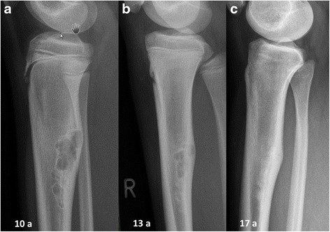
Original Image Producer: Herget et Al.. Credit: https://bmcmusculoskeletdisord.biomedcentral.com/articles/10.1186/s12891-016-1004-0. License: CC BY 4.0. Link to Source: https://commons.wikimedia.org/wiki/File:Fibroma_non_ossificante-RX.jpg .
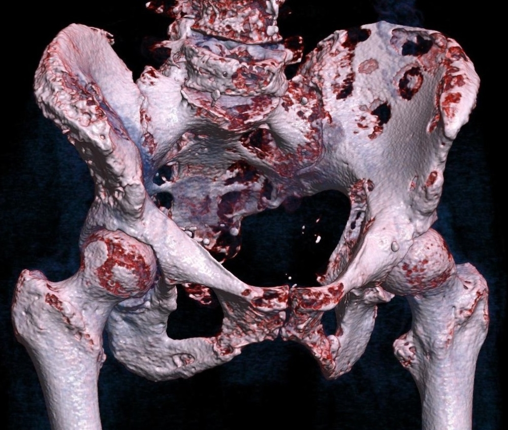
Original Image Producer: Mikael Häggström. Credit: Own work. License: CC0. Link to Source: https://commons.wikimedia.org/wiki/File:3D_rendered_CT_of_hip_bone_metastases.jpg .